- Mechanical Imaging and Tactile Imaging
- Prostate
- Breast
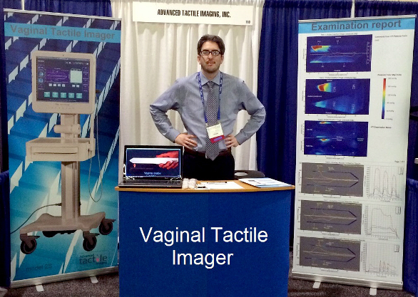
- Female Pelvic Floor
- Muscle
A significant part of the adult female population with the age are affected by at least one troublesome pelvic floor disorder such as pelvic organ prolapse, stres urinary urinary incontinence, pelvic pain, vaginal tissue athrophy, and sexual dysfunction. The female pelvic structures have varying viscoelastic characteristics across physiological states, healthy and diseased conditions.
The Vaginal Tactile Imager (VTI) obtains a high resolution mapping of pressures and assesses the strength of pelvic floor muscles within the vagina. The real time data as well the analysis information can then be viewed with an intention of assisting in the diagnosis and evaluation of vaginal and pelvic floor conditions. The VTI provides a large body of measurements to evaluate individual variations in support defects as well as identify specific potential markers to measure tissue properties and muscle function. The device is intended for use by physicians, surgeons and medically trained personnel.
The further progress in women's healthcare is possible if a patient with a problematic pelvic floor conditions could undergo opportunely imaging and biomechanical diagnostic tests; the results of which could be fed into a structured patient-specific diagnostic workflow to consider multiple treatment options and to suggest the optimal one for that patient.
Currently there are no efficient, standardized, non-invasive techniques for assessing of pelvic floor disorders and monitoring its treatment. VTI fills this gap providing a possibility to detect and quantitatively characterize the changes in physiological and anatomical manifestations of female pelvic floor transformation. The commercialization partner of Artann, a startup company Advanced Tactile Imaging. Inc., applies efforts to bring VTI to worldwide market.
Selected Publications and Patents
- Egorov V, van Raalte H, Lucente V, Sarvazyan A. Biomechanical characterization of the pelvic floor using tactile imaging. In: Biomechanics of the Female Pelvic Floor, Eds. Hoyte L, Damaser MS, 1st Editioin, Elsevier, March 22, 2016: 317-348.
- van Raalte H. Egorov V. Tactile imaging markers to characterize female pelvic floor conditions.Open Journal of Obstetrics and Gynecology 2015; 5: 505-515.
- van Raalte H, Lucente V, Egorov V. High definition pressure mapping of the pelvic floor muscles during Valsalva manever, voluntary muscle contraction and involuntary relaxation.Female Pelvic Medicine Reconstructive Surgery 2015; 21(5): S149-S150.
- Van Raalte H, Egorov V. Characterizing female pelvic floor conditions by tactile imaging.International Urogynecology Journal 2015; 26(4): 607-9, PMID: 25344223 with Video Supplement.
- Tactile imaging for quantifying vaginal elasticity in prolapse. Nature Reviews Urology 2012; 9(2): 60.
- Egorov V, van Raalte H, Lucente V. Quantifying vaginal tissue elasticity under normal and prolapse conditions by tactile imaging. International Urogynecology Journal 2012; 23(4):459-66.
- Egorov V, van Raalte H, Sarvazyan A. Vaginal Tactile Imaging. IEEE Transactions on Biomedical Engineering 2010; 57(7):1736-444.
- Egorov V, van Raalte H, Sarvazyan AP. Methods for assessment of pelvic organ conditions affecting the vagina. USA Patent 8,187,208; May 29, 2012.
- Egorov V, Sarvazyan AP. Methods for characterizing vaginal tissue elasticity. USA Patent 8,052,622; Nov 8, 2011